Fins-to-Limbs Transition: Apical fold morphogenesis and mesenchymal cell differentiation
Tohru Yano, Hiroki Yoshihara and Koji Tamura
Graduate School of Life Sciences, Tohoku University
One of morphological aspects of fins-to-limbs transition is loss of a fin-specific structure, fin ray, which includes back-to-back sheets of epidermis (apical fold, AF) and membrane bones (lepidotrichia). Studies suggesting similarities between fins and limbs have revealed shared mechanisms underlying their development, but little is known about development of the fin-specific structure.
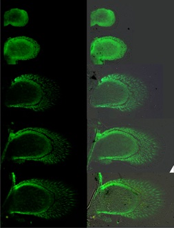
We studied some developmental features of the pectoral fin ray, focusing on AF morphogenesis/function and developmental origin of the fin ray mesenchyme. We found that the AF can anatomically and molecularly be divided into the proximal part (pAF) and the distal part (dAF) and that the dAF regulates AF outgrowth at later stages of fin development. Interestingly, removal of the whole AF did not result in fin truncation unlike AER removal in tetrapod limbs but gave rise to reformation of the AER and resultant elongation of the endosekeletal region.
Fin skeletons can be categorized into endochondral bones (endoskeletal elements) and membrane bones (fin ray elements). Endochondral bones in the pectoral fin correspond to limb skeletal elements in tetrapods that developmentally originate from the lateral plate mesoderm (LPM). On the other hand, the developmental origin of fin ray bones, fin-specific elements, remains unclear, although there are some arguments that the membrane bones are derived from neural crest cells migrating into the fins. Our cell lineage tracing with some labeling methods, however, provided evidence that those mesenchymal cells in the pectoral fin also originate from the LPM.